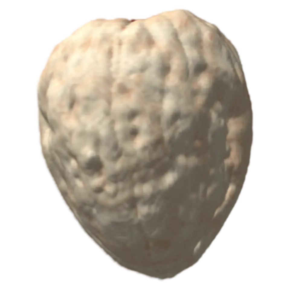
Volume Viewer. The same points, seen from the side. Drag down to look at it from above.
About 8 MB, on this tap only
Volume Viewer
A CT, MRI or microscope scan is a volume: a stack of slices, each a grid of numbers saying how dense or bright the object is there.
- Shelf
- Studio
- Kind
- Built from a recipe
- Made from
- Drag up or down on the volume to move the cut; tap it for the next preset. Play sweeps the cut through.
- Your own
- Your own volume
- Sound
- Yes
- Licenses
- CC BY 4.0, MIT
How to play
Drag up or down to cut into it; tap for the next window. Open your own scan in the Splash tab.
Its controls
| Control | Kind | Range | Start |
|---|---|---|---|
| Sweep the cut through (tap the volume for the next preset: bone, soft tissue, full range) | Tap | ||
| Sweep the cut through | Button | ||
| Next preset | Button | ||
| Volume | List | 3 choices | A walnut (X-ray CT) |
| View | List | 2 choices | 3D volume |
| Window | List | 4 choices | Full range (all but the air) |
| Level (your own window) | Setting | ||
| Width (your own window) | Setting | ||
| Colors | List | 5 choices | Bone and tissue |
| Opacity | List | 2 choices | Solid (everything in the window opaque) |
| Cut | List | 3 choices | Front to back |
| Only a thin slice at the cut | Setting | ||
| Voxel size in mm (blank: the file's) | Text |
About it
A CT, MRI or microscope scan is a volume: a stack of slices, each a grid of numbers saying how dense or bright the object is there. Each little block is a voxel. Slices are often farther apart than the pixels in them, so every voxel is placed at its real size.
The window picks which values show: below it is treated as air, and the colors run from its bottom to its top. Bone shows the densest parts, Soft tissue the range below them, Full range everything but the air (in a medical CT, in Hounsfield units: water 0, air -1000). Tap to step through them, or set your own. Drag to move the cut, or show one thin slice. Maximum intensity shows the brightest value along each line, as radiologists view blood vessels.
Open your own DICOM, NIfTI, TIFF stack or raw file in the Splash tab; it stays on your device. The samples, a walnut (CWI) and a young gar's head (Brian Metscher), are CC BY 4.0.
Details
- Your own files
- .dcm, .dicom, .ima, .nii, .gz, .zip, .tif, .tiff, .raw, .bin, .img, .vol, .dat
Sources and credits
| Work | By | License |
|---|---|---|
| Cone-Beam X-Ray CT Data Collection Designed for Machine Learning: Samples 1-8 (Walnut 1) | Henri Der Sarkissian, Felix Lucka, Maureen van Eijnatten, Giulia Colacicco, Sophia Bethany Coban, K. Joost Batenburg (CWI) | CC BY 4.0 |
| MicroCT images of 3 early gar (Lepisosteus osseus) stages between 12.7mm and 21.7mm (the 12.8 mm larva, averaged to 17.7 µm) | Brian Metscher (University of Vienna) | CC BY 4.0 |
Libraries
- MIT
- dicom-parser 1.8.21 (lane LSTUDIO, copied from Splashery)